New software UHasselt and ZEISS shows biomolecular movement in cells at the cutting edge
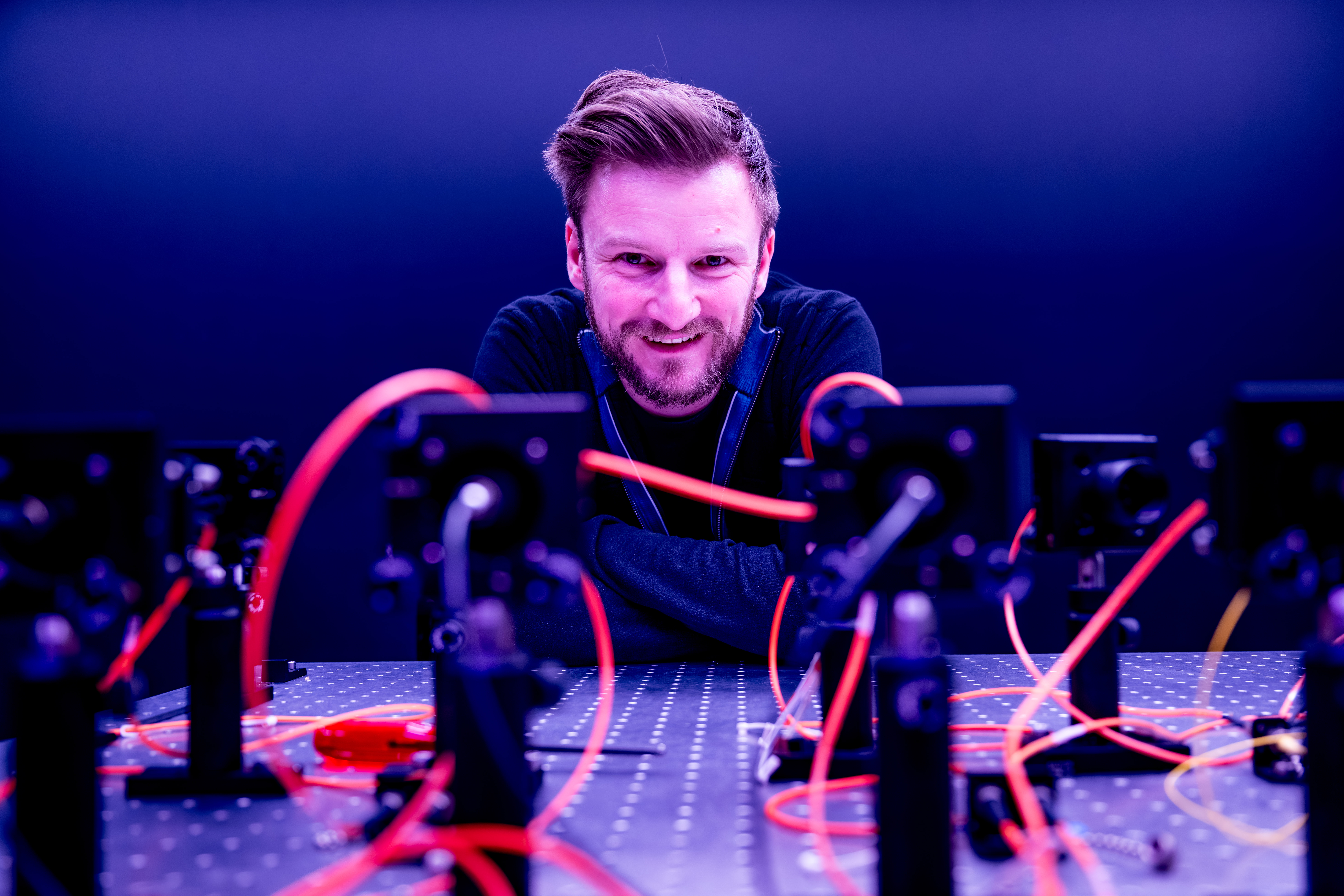
Researchers at BIOMED UHasselt, together with technology company ZEISS, which specializes in microscopes, developed a software module that dives deeper and better into human cells using confocal microscopy. This software can even be used to create so-called heatmaps, to see how biomolecules move within cells. "This will really boost the knowledge around cell biology and have an impact on a lot of biomedical and biological research," says Prof. Dr. Jelle Hendrix.


Software module
The software module from ZEISS and Prof. Dr. Jelle Hendrix's team, based on patented research from UHasselt and LMU Munich, can be used for confocal microscopy. In this type of microscopy, fluorescent dyes are used to stain biomolecules inside cells, the building blocks of the human body, and visualize them in 3D. By mathematically analyzing confocal videos of fluorescent biomolecules in living cells, researchers can infer, for example, how many fluorescent biomolecules are present, whether they interact with their environment, and whether the biomolecules form functional networks. Such information is crucial for understanding how cells work, but also for figuring out exactly what goes wrong in cells in certain diseases.
Great added value
"Over the years we have perfected these analyses, but still noticed that only niche researchers were employing them. The newly-developed software module now makes these analyses widely accessible, through its user-friendliness, by automating the most complex calculations, and by displaying the result of the analysis directly while sitting at the microscope," said Jelle Hendrix. "Whereas previously most confocal microscopists could only map the composition of the cells, the morphology, during their recording sessions, the new software module now allows them to simultaneously look inside the cell in razor-sharp detail, and see exactly which mechanisms are taking place. For example, heatmaps show in detail where and when certain biomolecules interact. A great added value within cell biology because knowledge about biomolecular contacts plays a key role in drug development, among other things."
Market leader
"ZEISS has been the market leader in this type of microscopy analysis for many years. The great added value of this software module is that the behavior of biomolecules is now not merely quantified, but also visualized. Microscopy images undergo a metamorphosis, so to speak, in which the functional behavior of biomolecules is displayed. In this respect, the module is truly unique," says Dr. Cicerone Tudor, Applications Specialist at ZEISS Benelux.
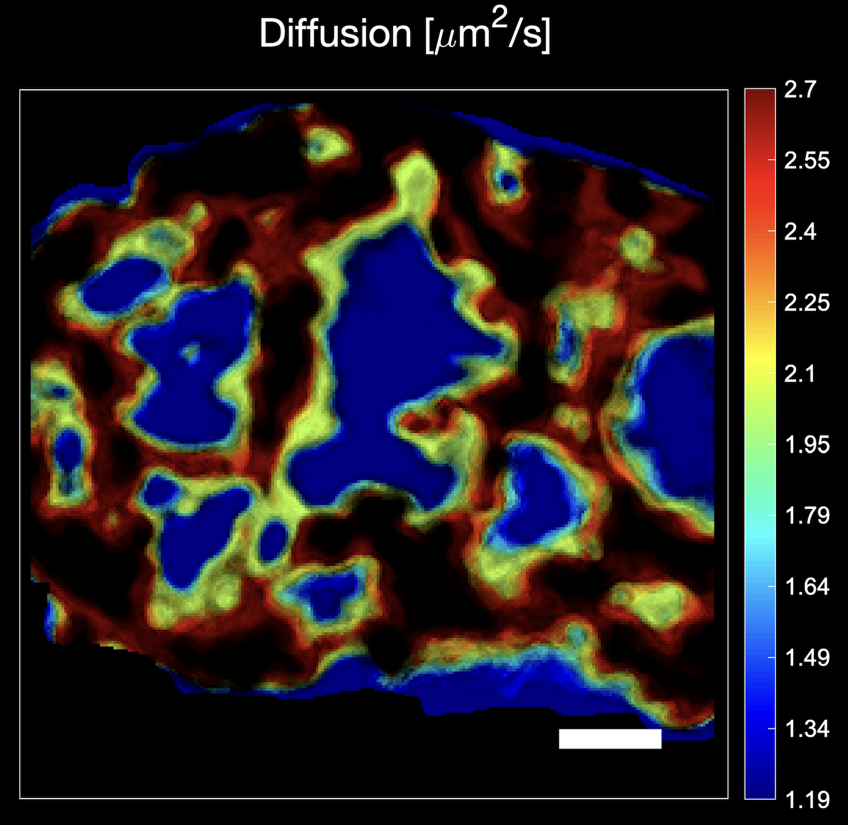
Image: Confocal microscopy heatmap indicating how fast biomolecules, in this case transcription factor LEDGF/p75 stained with green-fluorescent protein, move at different positions in the cell nucleus. White scale bar = 2 µm. Colored bar = diffusion coefficient in units of µm2/s.
Certificate of Excellence
The microscopy lab of the Biomedical Research Institute BIOMED at UHasselt also recently received the prestigious ZEISS Labs@Location certificate. A quality seal that the high-tech company ZEISS awards to laboratories that conduct research using various types of microscopy at the highest level. "Worldwide there are only about 30 laboratories that have this quality seal," says Prof. Dr. Jelle Hendrix. "This certificate is therefore a nice recognition, and it enables us to test out the latest technologies within microscopy in our lab and, such as now, develop new solutions and products together with ZEISS." Niek van Remortel, Head of Sales & Services at ZEISS Benelux: "The ZEISS Labs@Location label is a win-win because we can give potential customers demonstrations on top equipment at UHasselt. And this leads to more brand awareness for both parties."
Beautiful trajectory
The software from UHasselt and Zeiss has now been on sale for several weeks and will be rolled out in microscopy labs worldwide. "The process from idea to patent and the final development of the software was a very exciting collaboration between BIOMED researchers and business developers, the Tech Transfer Office of UHasselt, and the microscopy company ZEISS, and a nice example of knowledge transfer from the university to the outside world, one of the university's missions," says dr. An Voets, BIOMED's Industrial Research Fund Business Developer. "It's very nice to see that an idea we were playing with a few years ago has now taken its final shape. Especially now that we see that this can offer such added value within microscopy research," Jelle Hendrix concludes.